Figure 1
Partial SHOX duplications associated with various cases of congenital uterovaginal aplasia (MRKH syndrome): A tangible evidence but a puzzling mechanism
Daniel Guerrier* and Karine Morcel
Published: 24 March, 2021 | Volume 4 - Issue 1 | Pages: 001-008

Figure 1:
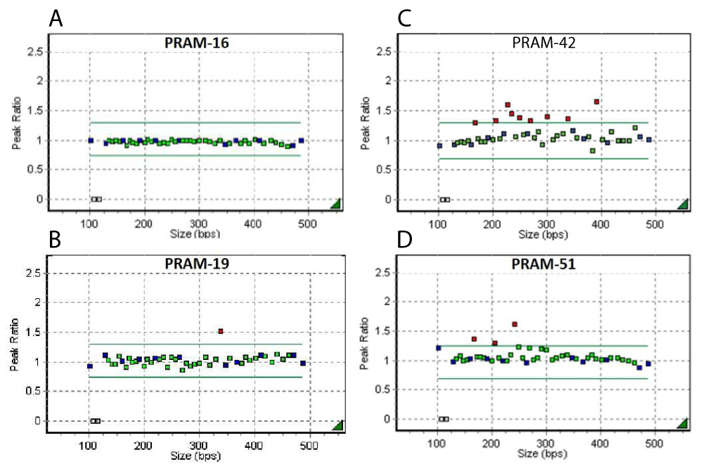
SHOX gene dosage in four independent MRKH patients (PRAM-16, -19, -42 and -51) assessed by the MLPA kit PO18-F1 SHOX . In the diagrams, MLPA probes are represented along the x-axis (size of PCR products) and the fluorescent intensity ratio is represented on the y-axis. Each probe is represented by a square (green for SHOX gene and surrounding regions, blue for internal controls, grey for the Y chromosome). The correspondence between the probes size and their respective location is shown on S1. The upper and lower arbitrary borders are shown respectively as a green upper and lower line. Probe ratios crossing the upper or the lower border are respectively indicative for a duplication or a deletion. Thus, a ratio of 1.5 (3:2) indicates the presence of an additional copy (heterozygous duplication) of a DNA stretch of a gene present in two copies in the genome, while a ratio of 0.5 (1:2) would indicate a heterozygous deletion. The detailed analysis of MLPA experiments is also included in S1. (A) Results of patient PRAM-16 showing no copy number variations. (B) Results of patient PRAM-19 showing a heterozygous duplication of 1 probed region (SHOX exon 5). (C) Results of patient PRAM-42 showing the duplication of 9 contiguous probed regions (4.7 kb upstream of the SHOX gene up to end of intron 6, 1.4 kb upstream of exon 7) and (D) Results of patient PRAM-51 showing the duplication of 3 contiguous probed regions (SHOX exons 1, 2 and 3).
Read Full Article HTML DOI: 10.29328/journal.jgmgt.1001006 Cite this Article Read Full Article PDF
More Images
Similar Articles
-
Partial SHOX duplications associated with various cases of congenital uterovaginal aplasia (MRKH syndrome): A tangible evidence but a puzzling mechanismDaniel Guerrier*,Karine Morcel. Partial SHOX duplications associated with various cases of congenital uterovaginal aplasia (MRKH syndrome): A tangible evidence but a puzzling mechanism. . 2021 doi: 10.29328/journal.jgmgt.1001006; 4: 001-008
Recently Viewed
-
Comparative characterization between autologous serum and platelet lysate under different temperatures and storage timesCamilo Osorio Florez*, Luis Campos, Jessica Guerra, Henrique Carneiro, Leandro Abreu, Andres Ortega, Fabiola Paes, Priscila Fantini, Renata de Pino Albuquerque Maranhão. Comparative characterization between autologous serum and platelet lysate under different temperatures and storage times. Insights Vet Sci. 2023: doi: 10.29328/journal.ivs.1001038; 7: 001-009
-
A Perspective on Conservation Technologies for Endangered Marine BirdsAnn Morrison*, Sonja Lukaszewicz. A Perspective on Conservation Technologies for Endangered Marine Birds. Insights Vet Sci. 2023: doi: 10.29328/journal.ivs.1001039; 7: 010-014
-
Intrauterine Therapy with Platelet-Rich Plasma for Persistent Breeding-Induced Endometritis in Mares: A ReviewThiago Magalhães Resende*,Renata Albuquerque de Pino Maranhão,Ana Luisa Soares de Miranda,Lorenzo GTM Segabinazzi,Priscila Fantini. Intrauterine Therapy with Platelet-Rich Plasma for Persistent Breeding-Induced Endometritis in Mares: A Review. Insights Vet Sci. 2024: doi: 10.29328/journal.ivs.1001045; 8: 039-047
-
Comparing Immunity Elicited by Feedback and Titered Viral Inoculation against PEDV in SwineMaría Elena Trujillo Ortega,Selene Fernández Hernández,Montserrat Elemi García Hernández,Rolando Beltrán Figueroa,Francisco Martínez Castañeda,Claudia Itzel Vergara Zermeño,Sofía Lizeth Alcaráz Estrada,Elein Hernández Trujillo,Rosa Elena Sarmiento Silva*. Comparing Immunity Elicited by Feedback and Titered Viral Inoculation against PEDV in Swine. Insights Vet Sci. 2024: doi: 10.29328/journal.ivs.1001044; 8: 028-038
-
Comparative Analysis of Water Wells and Tap Water: Case Study from Lebanon, Baalbeck RegionChaden Moussa Haidar, Ali Awad, Walaa Diab, Farah Kanj, Hassan Younes, Ali Yaacoub, Marwa Rammal, Alaa Hamze. Comparative Analysis of Water Wells and Tap Water: Case Study from Lebanon, Baalbeck Region. Insights Vet Sci. 2024: doi: 10.29328/journal.ivs.1001043; 8: 018-027
Most Viewed
-
Impact of Latex Sensitization on Asthma and Rhinitis Progression: A Study at Abidjan-Cocody University Hospital - Côte d’Ivoire (Progression of Asthma and Rhinitis related to Latex Sensitization)Dasse Sery Romuald*, KL Siransy, N Koffi, RO Yeboah, EK Nguessan, HA Adou, VP Goran-Kouacou, AU Assi, JY Seri, S Moussa, D Oura, CL Memel, H Koya, E Atoukoula. Impact of Latex Sensitization on Asthma and Rhinitis Progression: A Study at Abidjan-Cocody University Hospital - Côte d’Ivoire (Progression of Asthma and Rhinitis related to Latex Sensitization). Arch Asthma Allergy Immunol. 2024 doi: 10.29328/journal.aaai.1001035; 8: 007-012
-
Causal Link between Human Blood Metabolites and Asthma: An Investigation Using Mendelian RandomizationYong-Qing Zhu, Xiao-Yan Meng, Jing-Hua Yang*. Causal Link between Human Blood Metabolites and Asthma: An Investigation Using Mendelian Randomization. Arch Asthma Allergy Immunol. 2023 doi: 10.29328/journal.aaai.1001032; 7: 012-022
-
An algorithm to safely manage oral food challenge in an office-based setting for children with multiple food allergiesNathalie Cottel,Aïcha Dieme,Véronique Orcel,Yannick Chantran,Mélisande Bourgoin-Heck,Jocelyne Just. An algorithm to safely manage oral food challenge in an office-based setting for children with multiple food allergies. Arch Asthma Allergy Immunol. 2021 doi: 10.29328/journal.aaai.1001027; 5: 030-037
-
Snow white: an allergic girl?Oreste Vittore Brenna*. Snow white: an allergic girl?. Arch Asthma Allergy Immunol. 2022 doi: 10.29328/journal.aaai.1001029; 6: 001-002
-
Cytokine intoxication as a model of cell apoptosis and predict of schizophrenia - like affective disordersElena Viktorovna Drozdova*. Cytokine intoxication as a model of cell apoptosis and predict of schizophrenia - like affective disorders. Arch Asthma Allergy Immunol. 2021 doi: 10.29328/journal.aaai.1001028; 5: 038-040

If you are already a member of our network and need to keep track of any developments regarding a question you have already submitted, click "take me to my Query."


